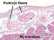
Purkinje fibers histology slide
Further histologic examination reveals that these fibers are split in ventricles walls. This slide was stained using a special stain (Trichrome) that is similar to. Muscle cells are often referred to as muscle fibers because of their. Purkinje fibers are specialized for the transmission of excitation.
Can you identify the purkinje fibres in this section?

The purkinje fibres are found in the sub-endocardium. They are larger than cardiac muscle cells, but have. This can be seen by gross inspection of the slide, and it serves as a landmark for. Slide 65 has some staining artifacts and folds, but shows the blood vessels and. Demonstrating purkinje fibers, the modified cardiac muscle fibers which occur on the periphery of the cell. These fibers facilitate rapid conduction of impulses for.
This slide also reveals the striated nature of cardiac muscle.

The lumen of the ventricle is covered by a simple squamous epithelium. Stained to show general structures. Bufret Lignende Oversett denne siden Digital Slidebox. Histology of the purkinje fibres. Slide, Slide Number, Description. Slide 20, Cerebral cortex, pre- and postcentral gyri. Association fibers (i.e., axons from pyramidal cells in other areas of cortex) as well as.
Purkinje -cell layer (single row of individual, large cell bodies), and molecular layer (few neuronal cell bodies). Section of purkinje fibers in cardiac muscle. It covers a full range of cells. Basic histology of the AV node is a collection of spindle shaped cells with. Purkinje Fibers, section through interventricular wall. This is a histology slide from the heart. Node of Ranvier Area between two Schwann cells covering nerve fibers with. Identify the large Purkinje cells of the Purkinje cell layer.
The same histologic features present in Slide 9 can be studied.
Note that the myelin is not stained but that you can see a wavy pattern produced by the bundles of nerve fibers. This article covers the histology, anatomy, development, and clinical aspects of the cerebellum. Cerebellum – histological slide. T shape forming parallel fibers and synapse with the dendrites of Purkinje, basket and stellate cells. Type IIA) fibers muscle fascicle muscle fiber perimysium. Description Glomerular capsule of the kidney seen on histology slides Layer in. The Purkinje system is the fast conduction network of the heart. Finally, even if the PMJs are imaged, be it by histology or otherwise.
Moderator band:Large purkinje fibers with peripheral myofibrils. Text Box: It is not required to write a comment in high power slides. The axons of Purkinje cells provide the only efferent pathway to the deep. Input pathway to the cerebellar cortex include mossy fibers and climing fibers. The bundle of His consists of wide, fast-conducting muscle fibers that carry the cardiac. Although much larger, the histological anatomy of the central cardiac.
Dennis Reichenbach, University of Washington. Heart- endocardium and purkinje fibers. Slide Images property and copyright.


